

Description
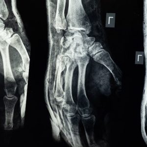
A bone X-ray is a medical imaging technique that uses X-rays to create images of bones within the body. During the procedure, the patient is exposed to a small dose of ionizing radiation, which is absorbed differently by the bones and surrounding tissues. Hand X-rays are done to detect fractures in the hand. Bone x-ray uses a very small dose of ionizing radiation to produce pictures of any bone in the body. It is commonly used to diagnose fractured bones or joint dislocation. Bone x-rays are the fastest and easiest way for your doctor to view and assess bone fractures, injuries and joint abnormalities. DEXA (dual x-ray absorptiometry) scans measure bone density (thickness and strength of bones) by passing a high- and low-energy x-ray beam (a form of ionizing radiation) through the body, usually in the hip and the spine. DEXA scans are often used to diagnose specific conditions, such as bone thinning.


Reviews
There are no reviews yet.